
by Leif Kullman
Increased diagnostic possibilities
by the use of the Internet
In my last report I tried to explain theoretically what a digital image is. And that is exactly the point, a digital image is rather theoretically, it exists in the computer (in the shape of a matrix system with 0 and 1 signs). But when we see the picture in the monitor it will be as an analogue image. When an object is radiographed, we get a continuous spectrum (all energy levels) of photons that hits our image medium (e.g. film). This "signal" is converted to a "digital image" by means of the analogue-digital converter in the computer. This digital image or matrix system can be manipulated in different ways. We can increase the number of grey levels in all the picture or in some defined areas of the picture (see figure 1). We can even make softwares that will ask the computer to find a place in the image where a caries lesion is situated. The computer is prompted to look for an area in the image, where the grey level is of a defined, "caries" level. When we have finished our image manipulation and want to see the picture in our monitor, the analogue-digital converter must work again, dumping the picture in a continuos scale (analogue format) on the screen again. Unfortunately our monitors today only have a capacity to use around 64 grey levels out of our 256 possible.
Original intraoral
radiograph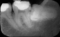 |
|
A small area of the
original radiograph is selected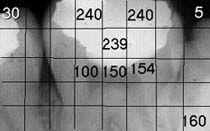 |
The
principle of transforming an analogue image to a digital. Each pixel, there are |
This month I will report on why I
believe that it will be increased possibilities for diagnosing pathology in the future by
means of computers and Internet. With this statement I don't mean that the future
specialist in Oral Radiology will be more clever than the older ones. Perhaps the opposite
will happen, future specialist can become more addicted to our advanced equipment and be
totally handicapped without them. No, the great advantage with our IT world will be that
so many dentist will be able to increase their diagnostic possibilities by means of
Internet. This is not as difficult to understand as the difference between analogue and
digital images was. Every clinically working Internet connected dentist will have the
gathered knowledge of the whole world
at his feet. If he find something strange in a radiograph and is unsure about the
diagnosis he can send it to a specialist that is connected to the net. Probably he will
have the answer the same day (if it is possible to state it without PAD). Today many
specialists have started discussion groups in their field, where they discuss common job
matters. In Oral Radiology we have one with members from all the world, the Northamericans
being the most active. Within this group we have started to send radiographs helping each
other to diagnose.
Many institutions and Schools of Dentistry have their home pages, where they among other
things show interesting cases they have had. Anamnestic records, radiographs and other
relevant recordings are shown. If you are interested in one case, it is possible to copy
e.g. a radiograph and store it on your own computer. Finally there are also more advanced
diagnosing software on the net, with which it is possible to establish a diagnosis or
differential diagnosis. One of them is ORAD, which can be reached from the
homepage of ODIS. With this software it is possible to get a proposal to a diagnosis based
on the clinical, anamnestic and radiographic recordings. By selecting check boxes such as
age of patients, area for the pathology, appearance of the pathology and so on a final
diagnosis is proposed (or differential diagnosis).
Next month:
Next time we meet I will tell you a little more about advantages with digital imaging.
Leif Kullman