
by Leif Kullman
Dear ODIS- reader, imagine the following scenario:
When you arrive to your clinic in the morning you turn on your computer. After you have answered with your password the computer finds one mail for you from a friend on the other side of earth - Australia. He was yesterday on a picnic with his family and took some pictures with his new digital camera, when they were bathing. When he came home he could look at these pictures in his PC and attach them to an e-mail sent to you. Now you have them on your screen. You smile when you see your friend and his family in the sunshine, outside your own window the first snowflakes for the season are falling down. You think that our world has become very small.
After you have had your first patient you sit down at the computer again to order some impression materials from your dental supplier. You connect to them by means of Internet and can directly in their homepage look at the things that you need to buy. You make your choices and know that you will have the things the day after delivered to your practice.
In the afternoon you have an urgent patient. The patient is a young healthy girl, 9 years old. She and her mother told you that she has had no pain, but has noticed a swelling on the right side of the mandible during some weeks. You have recently bought a new digital panoramic equipment and the nurse takes the panoramic down under.
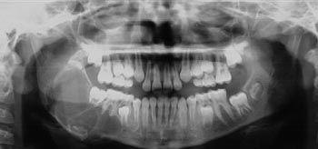
You are not sure of the diagnosis in this case and are in a hurry, the lesion seems very large. You e-mail to your friend who is a specialist in oral radiology and ask for his opinion.
During lunch time you have a couple of minutes free. You start your mail program again to see if some new messages have arrived since the morning. Besides of your mentioned friend, you are also a member of a mailing list, discussing new materials in odontology. Most of the members are working in the U.S.A and often they have tipped you about advantages and shortcomings with new materials. Today there are one member who has started to use a new intraoral radiographic film and since the beginning he has had problem with dark horizontal lines across the developed film. He suspects that it depends on his automatic developer, which damage the film emulation and now he wonders if someone else has met the same problem. You have had about the same problem from some other colleagues and you think that in the future when all are using digital equipment's these kind of film problems will not exist.
Toward a preliminary diagnosis
Two hours later, having your afternoon coffee, you get an answer from your radiologist. The answer describes a multiple radioluscent area, in the mandible and ramus ascendens of the right side. The border is well defined and the outer compact bone is deviating inferior. He suggests the following differential diagnosis based upon the radiographic appearance: Ameloblastoma, aneurysmal bone cyst or keratocyst. He also states that with all probability the lesion is benign. You can call the patient and give her a new appointment time.
Dear reader, in a near future I think this will be a reality and I look forward to it.
Next month I will tell you more about panoramic equipment, including some comments about the new digital units.
Leif Kullman