
by Leif Kullman
A routine case with an interesting appearance in the radiograph.
This month we are going to look at a patient with a pathological lesion, which is rather frequent seen in the jaws (comprises about 5% to 15 % of all cysts in the jaws).
A cystic lesion or something else?
Our patient is a middle-aged woman, completely healthy. She has no symptoms from the jaws or teeth. During her annual visit to her general private practitioner the following
radiographs is taken
Fig.1.
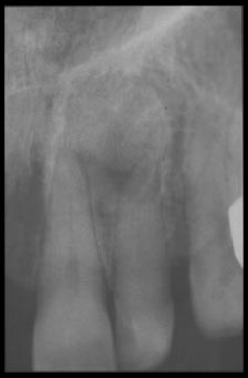
In regio 21,22 an area can be seen with a changed bone structure. The area is mostly well bordered and has a size of about 1 cm. No tooth displacement can be seen. The lesion looks mostly unilocular. No resorption can be seen in the adjacent teeth. Since the general practitioner is uncertain about the diagnosis she contacts me as a specialist in Oral Radiology to discuss possible differential diagnosis. The radiographic signs are not easy to evaluate. Some anatomical deviation may exist in the region and explain the appearance but this diagnosis seems unlikely. The borders of the area are in some places rather irregular but mostly well defined and this indicate that some kind of a benign tumor could not be excluded as a possible diagnosis, for example an early periapical cementdysplasia. Either can an early ameloblastoma be eliminated as a diagnosis. However the mainly hard tissue border indicates also that
some kink of cyst can be the probable diagnosis. For example an odontogenic keratocyst. Finally a giant cell granuloma had to be included in probable diagnosis. Since all radiographic signs indicated a benign lesion, and as the patient had no pain or symptoms at all, we decided to wait a year.
The following year was the patient still free from symptoms and a new radiograph looked like this:
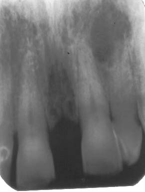
Fig 2.
The lesion had increased in size and became more radiolucent and we could exclude the anatomical abberation as a diagnosis (and could now name it a lesion). It was therefore decided to refer the patient for a surgical exploration. The surgeon found a cavity with a cystic epithelium during his operation, containing a semifluid, thick cheesy material. When the report came from the Oral Pathology department, the diagnosis was odontogenic keratocyst. In a textbook in Oral Radiology (Oral Radiology.Principles and Interpretation by Goaz P.W. and White S.W. Third Edition. Mosby.1994.U.S.A.) the following can be read about odontogenic keratocysts: " this cyst is frequently
indistinguishable radiographically from any other odontogenic cyst. The majority appear as radiolucencies in the mandible, with more than 90 % posterior to the canines. The size varies from small to 5 cm. Tooth displacement is common and the majority are unilocular with smooth borders."
The first year after the operation the lesion looked to heal, but after further one year the beginning of a new radiolucent are could be seen( figure 3) and a reoperation had to be performed.
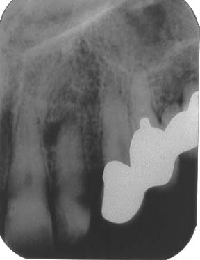
Fig. 3.
One of the most prominent characteristics of a keratocyst is its ability to recurrence. They should therefore be followed regularly up to
ten years after an operation.
Next month I will report about theradiographic characteristics
on a malign lesion i the jaws.