
by Leif Kullman
Radiology in oral implant therapy.
Twenty-three years ago when I
graduated as a dentist, there was on principle only one therapy alternative for toothless
patients, namely a full denture. Sometimes you could see, so called subperiostal implants,
often with radioluscenses in the surrounding bone, indicating inflammation. The biological
acceptance of our body was small for these kinds of implant. Today we have a quite
opposite situation, anyway in Sweden. Many toothless patients get implants instead of
dentures, which become osseointegrated with the bone. Most of these are made of titanium.
My report this month will focus on radiographic methods in implant therapy.
Three different situations can be recognized, when radiographs are indicated, namely the presurgical investigations and investigations during installation and finally during the post-installation controls.
Presurgical investigations
Besides, as always when we take radiographs, to be able to diagnose possible pathology
and the usual anatomic details in the jaws presurgical radiographs should give a
possibility to perform accurate measurements. Preferably also give a possibility to
identify the location of the image relative to fixture site. Additionally it is useful if
they can give a cross-sectional view of the dental arch, to visualize the inclination of
the alveolar process at the future implant site and the height.
Finally a great advantage is an ability to evaluate the density of bone (the bone quality).
The following modalities are or may become useful:
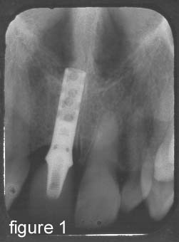
Intraoral radiographs: Pathology may be diagnosed and measurements may be performed with an ordinary ruler, since the magnification is small (figure 1).
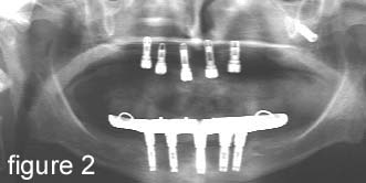
 Panoramic radiographs: May be used too, instead of intraoral
radiographs (or as a complement). However a magnification must be considered, which is of
different size for different panoramic equipments. One of the great advantages with this
method is that a whole jaw or both jaws may be visualized in one image, however there are
not the same sharpness and resolution in panoramic images (figure 2) as in intraoral
radiographs.
Panoramic radiographs: May be used too, instead of intraoral
radiographs (or as a complement). However a magnification must be considered, which is of
different size for different panoramic equipments. One of the great advantages with this
method is that a whole jaw or both jaws may be visualized in one image, however there are
not the same sharpness and resolution in panoramic images (figure 2) as in intraoral
radiographs.
Lateral or profile images: Useful to see above all the relation between the alveolar
crists in lower and upper jaw. But can also be used to roughly evaluate the total height
of the frontal parts of the jaws.
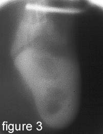 Conventional tomography (figure 3). This method is recommended
over OPG with planned mandibular implants posterior of foramen mentale. It is possible to
get a more reliable and more accurate image than with OPG and the facial-lingual space can
be viewed. However the precise location of the image slices are sometimes difficult to get
but some sort of guide to over-come this can be used.
Conventional tomography (figure 3). This method is recommended
over OPG with planned mandibular implants posterior of foramen mentale. It is possible to
get a more reliable and more accurate image than with OPG and the facial-lingual space can
be viewed. However the precise location of the image slices are sometimes difficult to get
but some sort of guide to over-come this can be used.
Figure 3. Tomographic image from the
lower jaw
Computed tomography: This is the best method to understand the precise configuration or topography of the jaws. It is also possible to make exact measurements of the height, width of the alveolar bone of the jaws. The size of a future implant can therefore reliable be chosen. Both linear and angular measurements may be performed.
Magnetic Resonance imaging (MRI) This is a computerised tomographic method yielding images with excellent spatial resolution. The soft tissues are also well depicted and the patient is preserved from usual x-ray radiation. However, the soft tissues are of minor importance in implant radiology, but the method may be useful to study the healing process when bone grafts are used. One great advantage is freedom of special slice position and angulation of the patient with this method. Disadvantages are: No signal from the cortical bone and an expensive method!
During installation
In principle, intraoral and panoramic images are the most used here, for example to check
the position of a fixture before the distance operation.
Postinstallation controls
During this phase, intraoral and panoramic radiographs are also the most used methods.
However if bone transplants have been installed prior to the implant installation, CT (or
MRI) may be useful to check if this bone has been successfully integrated with the
ordinary alveolar bone of the patient.
Next month I will discuss some health physics matters, particular how these matters often are described in daily newspapers.